- Teacher: Krista Lewis
- Teacher: Chelsea Muckey
Available courses
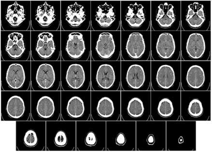
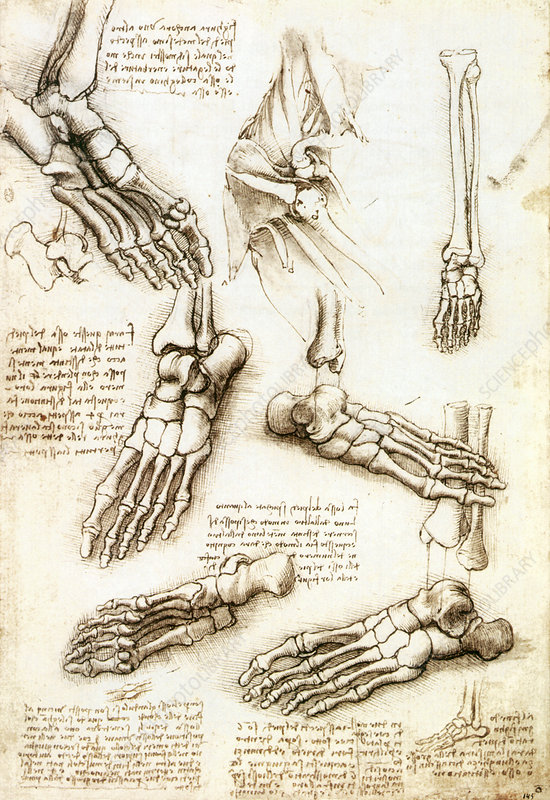
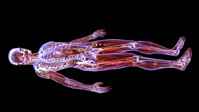
Study is made of human anatomy and its function. Areas include general cell and tissue
structure, the respiratory system, general bone structure, the lower extremities, the upper
extremities, and the genitourinary system. Detailed topographic anatomy, positioning
landmarks, and radiographic appearance are covered.
- Teacher: Krista Lewis
- Teacher: Chelsea Muckey
A continuous process beginning with supervised correlation of theory and actual
performance of examinations to unassisted performance and decision-making in a variety
of situations. Clinical rotation guidelines assist the student as they progress form one
area to another. Regular evaluations are an integral part of this process.
- Teacher: Krista Lewis
- Teacher: Chelsea Muckey
The course encompasses subjects pertinent to the care and examination of the patient and
work within the profession. Study is made of patient assessment and communication,
safety, infection control, vital signs, pediatric imaging, and geriatric imaging.
- Teacher: Krista Lewis
- Teacher: Chelsea Muckey
This course investigates detection and measurement of radiation, sources of radiation
exposure, occupational and general public dose equivalent limits, methods for
minimizing exposure to patient, self, and others, and application of federal and state
regulations.
- Teacher: Robert Hughes
This course introduces students to the factors that control the radiation exposure to the
image receptor. The primary exposure factors, differential absorption, patient and
equipment factors that affect exposure, spatial resolution, distortion, and magnification
are covered. Students will work to formulate a technique chart. Emphasis is placed on
practical applications of basic laws and theories in problem-solving situations and
formulation of techniques in the production of quality images.
- Teacher: Robert Hughes
Study of the common and supplementary positions and procedures for the respiratory
system, abdominal cavity, lower extremities from the knee to the toe, and upper
extremities from the elbow to the finger. Anatomical landmarks, anatomy, body habiti,
and special patient considerations are emphasized. Laboratory demonstration is used for
patient correlation.
A continuous process beginning with supervised
correlation of theory and actual performance of examinations to unassisted
performance and decision-making in a variety of situations. Clinical rotation guidelines assist the
student as they progress form one area to another. Regular evaluations are an integral part of
this process.
- Teacher: Robert Hughes
- Teacher: Krista Lewis
- Teacher: Chelsea Muckey
Study is made of the basic structure of matter,
electricity, magnetism, and their interrelationships. That knowledge is applied to the construction
and function of x-ray generators, circuitry components, the x-ray tube, and
fluoroscopic image intensifier. X-ray
production and emission are discussed in detail.
- Teacher: Robert Hughes
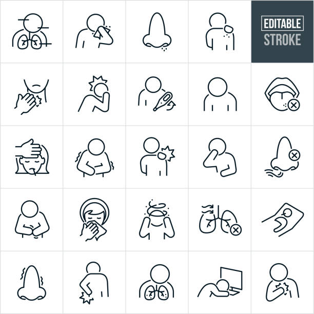
Discussion is made of medical/surgical diseases with diagnostic evaluation and treatment. The impact of disease and pathology is explored in patient evaluation, image quality, and performance of various radiographic procedures. Areas include the imaging modalities, general pathological processes, the respiratory system, and the portions of the skeletal system.
- Teacher: Krista Lewis
The course encompasses subjects pertinent to the care and examination of the patient and work within the profession. Study is made of medical emergencies, trauma, urologic and gastrointestinal procedures, aseptic technique, ECG, and an introduction to pharmacology and drug administration.
- Teacher: Krista Lewis
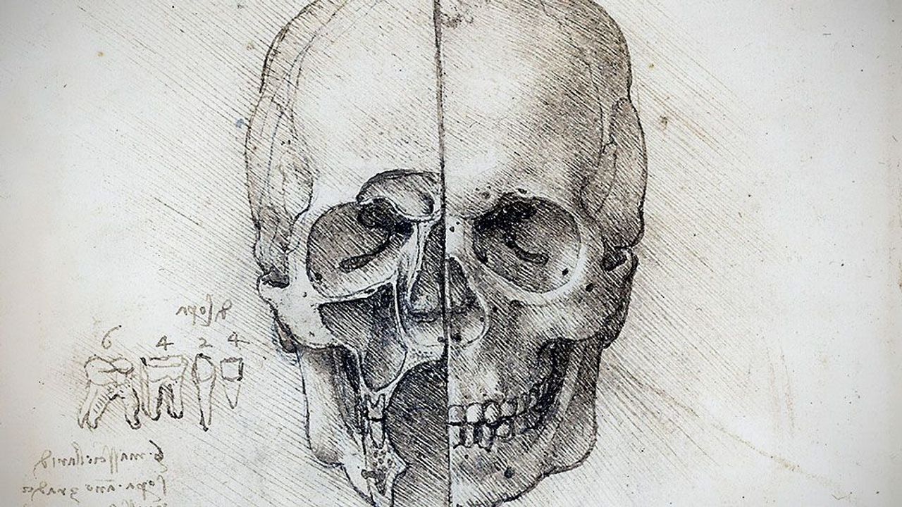
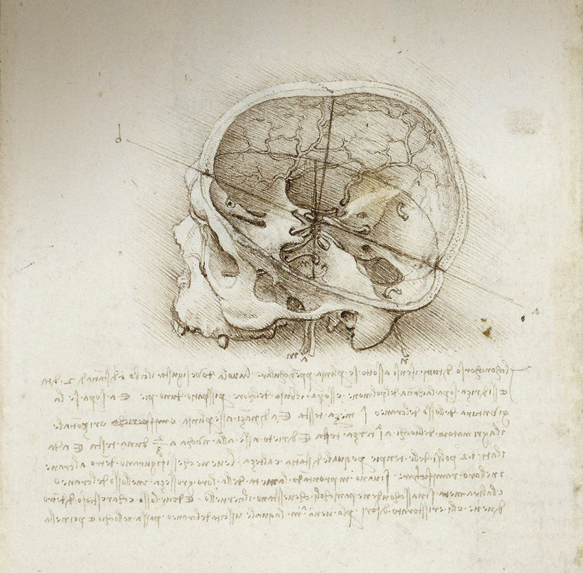
Study is made of human anatomy and its
function. Areas include the
gastrointestinal system and accessory organs, spine and thorax, skull, heart,
and cardiovascular system. Detailed
topographic anatomy, positioning landmarks, and radiographic appearance are
covered.
- Teacher: Krista Lewis
Study of the common and supplementary positions
and procedures for the upper and lower GI system, urinary system, pelvic girdle
and femur, and shoulder girdle and humerus.
Anatomical landmarks, anatomy, body habiti, and special patient
considerations are emphasized.
Laboratory demonstration is used for patient correlation.
- Teacher: Robert Hughes
- Teacher: Krista Lewis
- Teacher: Chelsea Muckey
This course involves lectures and practice in specialized procedures and interventional exams. Study is made of positioning, supplies, precautions, and patient care during these procedures. Topics include myelograms, hysterograms, arthrograms, and general tomography.
- Teacher: Robert Hughes
- Teacher: Krista Lewis
- Teacher: Chelsea Muckey
A continuous process beginning with supervised correlation of theory and actual performance of examinations to unassisted performance and decision-making in a variety of situations. Clinical rotation guidelines assist the student as they progress form one area to another. Regular evaluations are an integral part of this process.
- Teacher: Krista Lewis
- Teacher: Chelsea Muckey
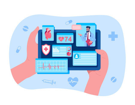
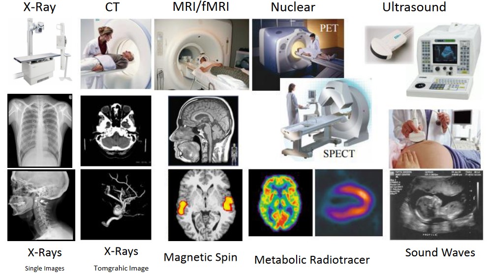
The student is introduced to the equipment and general operation of the imaging and therapeutic modalities. Topics include tomography, DEXA, MRI, CVIT, mammography, sonography, nuclear medicine, radiation therapy, and dosimetry.
- Teacher: Krista Lewis
Study is made of the role of digital systems in the modern imaging environment. Correlation of basic exposure principles as applied to the creation, pre-processing, post-processing, display, and storage of images in a digital environment. Special consideration is given to the differences in CR and DR as well as the quality factors of the display system.
- Teacher: Robert Hughes
This course relates the physical and biologic effects of radiation to human life and the environment. Interactions of x-rays and matter are studied in depth. Effects on a molecular, cellular, and whole body level are correlated to short and long term effects. Survival statistics and risk estimates are also studied.
- Teacher: Robert Hughes
Study of the common and supplementary positions and procedures for the spinal column, bony thorax, and skull. Anatomical landmarks, anatomy, body habiti, and special patient considerations are emphasized. Laboratory demonstration is used for patient correlation.
- Teacher: Robert Hughes
- Teacher: Krista Lewis
- Teacher: Chelsea Muckey
Discussion is made of medical/surgical diseases
with diagnostic evaluation and treatment.
The impact of disease and pathology is explored in patient evaluation,
image quality, and performance of various radiographic procedures. Areas include portions of the skeletal system
and the digestive system.
- Teacher: Krista Lewis

- Teacher: Krista Lewis
- Teacher: Robert Hughes
- Teacher: Krista Lewis
- Teacher: Chelsea Muckey
- Teacher: Krista Lewis
- Teacher: Chelsea Muckey
- Teacher: Krista Lewis
- Teacher: Krista Lewis
Students prepare a research paper and scientific exhibit from a topic within the radiation sciences. This course provides practical skills and steps needed in the planning, design, collection of data, analysis, and inference of findings for the research process. Students will also practice the practical writing of departmental policies and communications.
- Teacher: Robert Hughes
This course involves lectures and practice in specialized procedures and interventional exams. Study is made of positioning, supplies, precautions, and patient care during these procedures. Topics include ERCP, Sialograms, Lymphangiograms, Venograms, surgical radiography, and trauma radiography.
- Teacher: Krista Lewis
- Teacher: Chelsea Muckey
- Teacher: Chelsea Muckey


- Teacher: Robert Hughes
- Teacher: Krista Lewis
- Teacher: Chelsea Muckey
Assisting the transition out of an academic environment,
this course incorporates academic reviews, certification preparation, and
professional preparation with a focus on critical thinking and problem solving
skills in a professional environment.
Correlation is made between larger health care concerns and factors
impacting the profession.
- Teacher: Robert Hughes
- Teacher: Robert Hughes
- Teacher: Krista Lewis
- Teacher: Chelsea Muckey
- Teacher: Robert Hughes
- Teacher: Krista Lewis
- Teacher: Chelsea Muckey
- Teacher: Robert Hughes
- Teacher: Krista Lewis
- Teacher: Chelsea Muckey
Holding place for all posted lecture and reference materials.
- Teacher: Krista Lewis
- Teacher: Krista Lewis